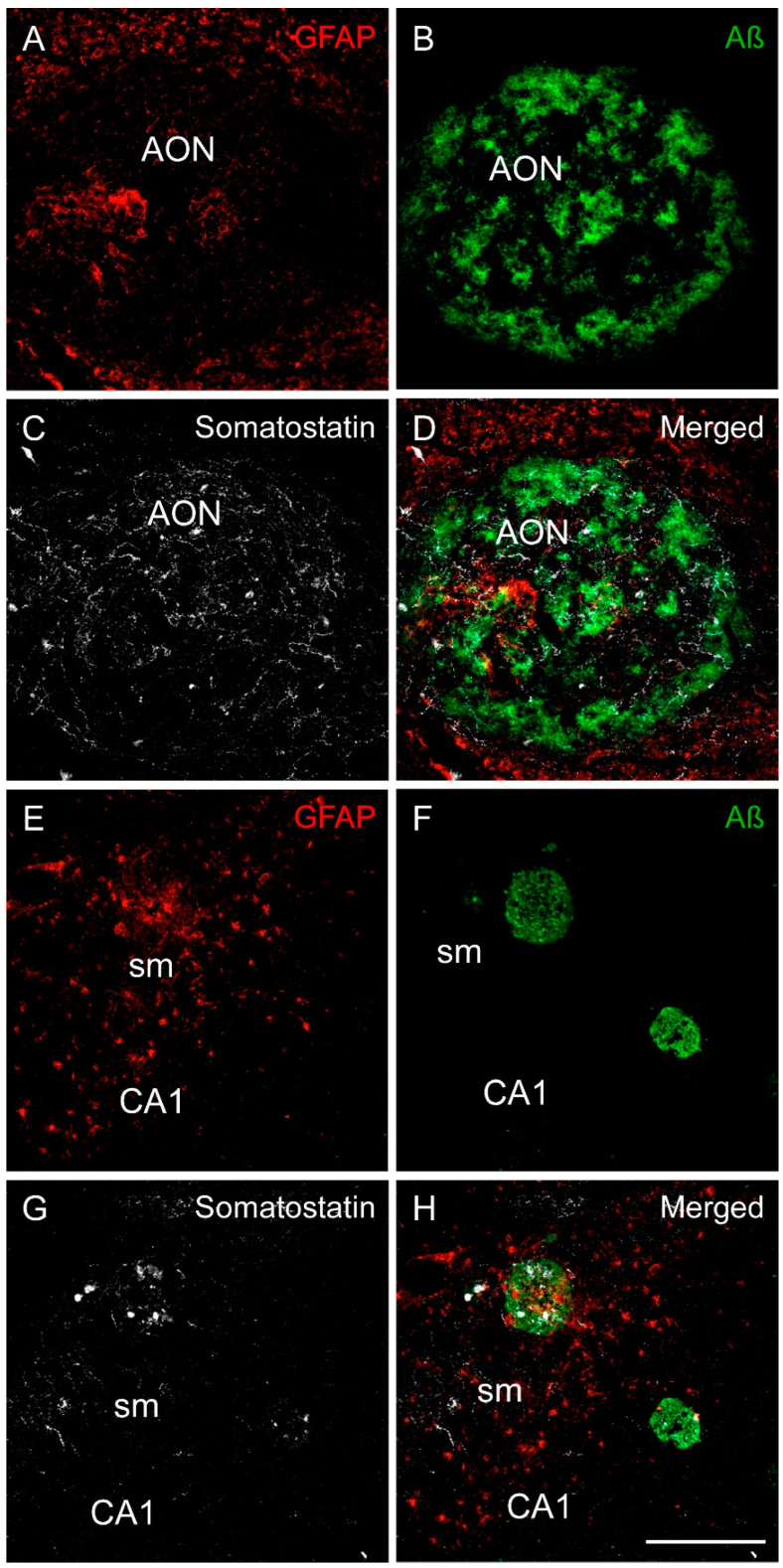
Figure 7.
Immunofluorescence staining of somatostatin, amyloid-β and GFAP in the human olfactory limbic systems in AD. Representative images show staining for GFAP (astrocytes in red), amyloid-β (green) and somatostatin (white) in the anterior olfactory nucleus (A–D) and in the CA1 region of the hippocampus (E–H). In the anterior olfactory nucleus, there were less astrocytes than in the surrounding area (A), whereas amyloid-β (B) and somatostatin (C) were tightly coexpressed (D). In the hippocampus, astrocytes were abundant in the molecular layer of CA1 (E). In addition, astrocytes, amyloid-β plaques (F) and somatostatin (G) were commonly coexpressed in this area (H). Scale bar A–H 200 µm.

