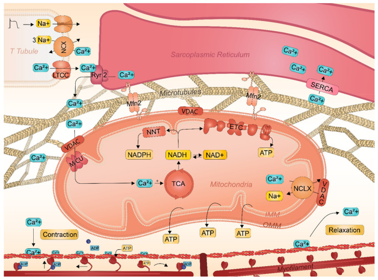
Figure 2.
Schematic representation of Ca2+ transient handling and contraction in atrial cardiomyocytes and the role of SR and mitochondria. An action potential activates the Ca2+ channels in the T-tubule, resulting in an influx of Ca2+ into the cardiomyocytes. Subsequently, Ca2+ crosses the OMM through VDAC and is transported into the mitochondrial matrix via the MCU. Ca2+ efflux from the matrix is mediated by the NCLX. Cytosolic Ca2+ binds to cardiac troponin-C, which moves the troponin away from the actin binding site to form cross-bridges between actin and myosin. The myosin head binds to ATP and pulls the actin filaments towards the center of the sarcomere resulting in cardiomyocyte contraction. Dropping of the intracellular Ca2+ concentration returns the troponin complex to the resting position, thereby effectively inhibiting cardiac contraction and initiating relaxation.

