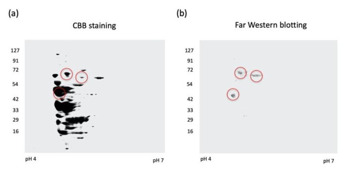
Figure 1.
Two-dimensional electrophoresis and Far-Western blotting. Bacterial cells of A. defectiva ATCC (7.5 × 10⁶ CFU) were lysed and isoelectric focusing, followed by SDS-PAGE, was applied. Gel stained with Coomassie Brilliant Blue (CBB) (a) and the membrane blotted protein was incubated at 4 °C overnight with 50 µg/mL of fibronectin and then probed with rabbit anti-fibronectin antibodies diluted at 1:500 in PBS (pH 7.0). The membrane was then washed with PBST and incubated with a 1:2500 dilution of goat anti-rabbit IgG conjugated with HRP (b).

