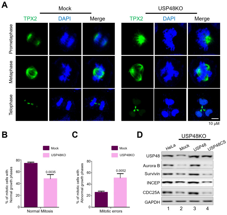
Figure 5.
USP48 regulates Aurora B function during mitosis. (A) The spindle assembly factors in mock (left panel) and USP48KO (right panel) HeLa cells were stained with TPX2 antibodies. DAPI was used to stain the nuclei. Scale bar = 10 μM. Mitotic defects were observed upon depletion of USP48. The graph represents the percentage of cells exhibiting (B) normal mitotic phases in mock (74 ± 2), and USP48KO (48 ± 7) and (C) abnormal mitotic phases in mock (26 ± 2) and USP48KO (51 ± 7.55) (n = 3, p values are represented on each graph). (D) USP48KO HeLa cells (lane 2) were transfected with either Flag-USP48 (lane 3) or Flag-USP48CS (lane 4) and compared with wild-type HeLa cells (lane 1). Transfected cells were synchronized with 100 ng/mL Nocodazole for 18 h prior and analyzed by Western blot against the indicated antibodies.

