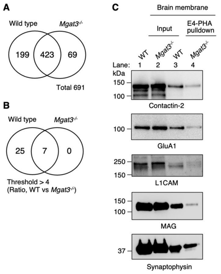
Figure 2.
Identification of bisecting GlcNAc-bearing N-glycosylation sites in the brain. (A) Venn diagram indicating the total number of peptides identified by LC-MS. (B) Venn diagram indicating the number of peptides confirmed to be N-glycosylated. (C) The representative results of Western blot analysis of brain membrane fractions incubated with E4-PHA lectin beads. Membranes were probed with anti-contactin-2, anti-α-amino-3-hydroxy-5-methylisoxazole-4-propionic acid (AMPA)-type glutamate receptor 1 (GluA1), anti-neural cell adhesion molecule L1 (L1CAM), anti-myelin-associated glycoprotein (MAG), and anti-synaptophysin antibodies.

