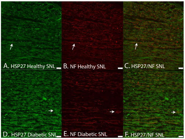
Figure 8.
Images demonstrating double staining HSP27 (arrows in left panels) and neurofilaments showing that HSP27 expression is presented in neurofilament-stained axons (middle panels) at the site of lesion (SNL) in healthy Wistar and diabetic GK rats. The images (A,B) are merged together in (C). The images (D,E) are merged together in (F). Bar = 100 μm.

