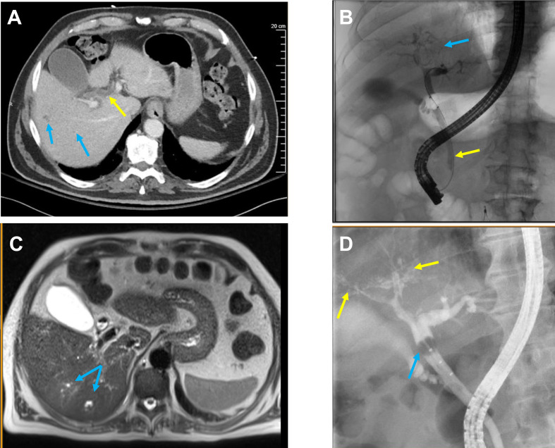
Figure 2.
Radiological imaging. (A) CT of the abdomen and pelvis with intravenous contrast taken day 51 from COVID-19 diagnosis demonstrating multiple subcentimetre low-density lesions in the right hepatic lobe (blue arrows) and dilatation of the common bile duct (yellow arrow). (B) Endoscopic retrograde cholangiopancreatography (ERCP) with intrahepatic radicals (blue arrow) and stent insertion (yellow arrow). (C) MRI of the abdomen day 164 from admission showing mild intrahepatic biliary ductal dilatation and mild patchy T2 hyperintensity in the right liver (blue arrow), possibly concerning for primary sclerosing cholangitis. Additionally, diffuse biliary hamartomas were visualised. (D) ERCP on day 150 with evidence of relative ductopenia of the right and left ducts with subtle irregularity and beading consistent with secondary sclerosing cholangitis (yellow arrow). A limited cholangiogram done during the procedure demonstrated a filling defect concerning for biliary casts (blue arrow).

