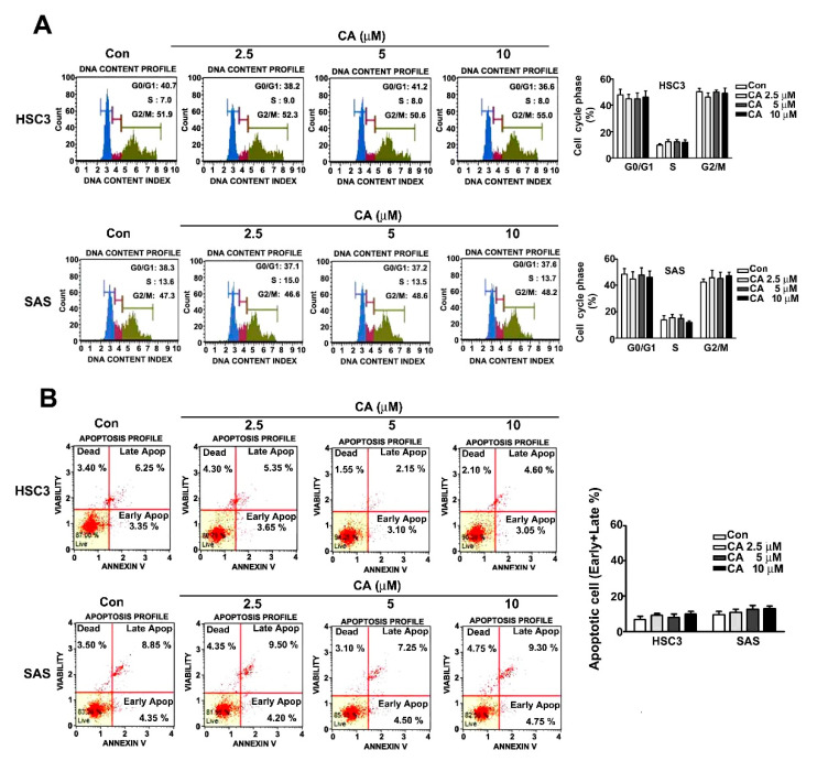
Figure 2.
Effect of CA on cell cycle arrest and apoptosis induction in human HSC3 and SAS OSCC cells. (A) The regulation of cell cycle distribution was analysed using PI staining by flow cytometry. (B) Apoptosis induction in human HSC3 and SAS cells exposed to various concentrations (0, 2.5, 5 and 10 μM) of CA by Annexin V/PI staining through flow cytometry. (Mean ± SE, n = 3). Control: untreated cells.

