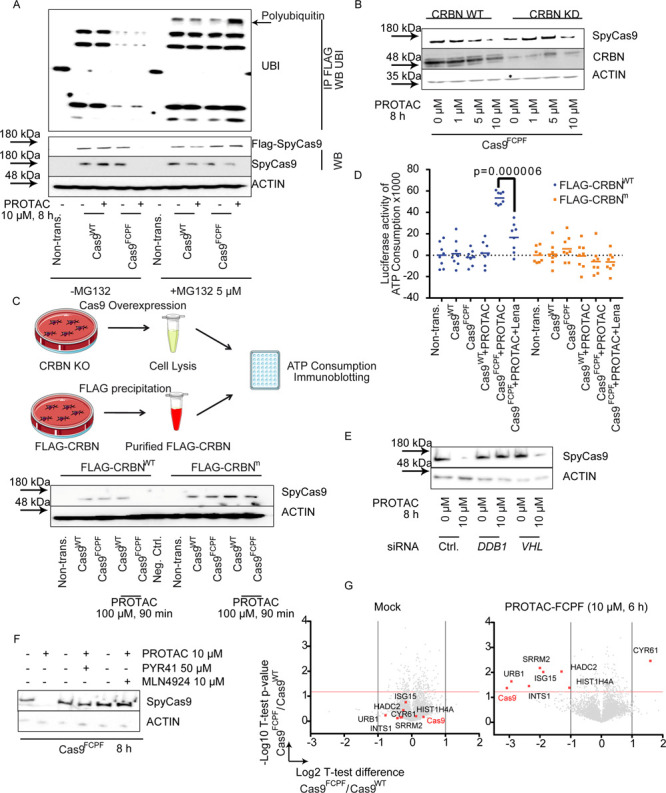
Figure 3.

The PROTAC-FCPF-induced Cas9FCPFprotein degradation is ubiquitin-dependent. (A) MG132 inhibits PROTAC-FCPF-mediated Cas9FCPF protein degradation. HeLa cells expressing Cas9WT or Cas9FCPF were treated with PROTAC-FCPF (10 μM) in the absence or presence of MG132 (5 μM) for 8 h. DMSO was used as a control. Cas9 protein was immunoprecipitated, and immunoblotting was performed for detecting the ubiquitination. (B) Comparison of PROTAC-FCPF-induced Cas9FCPF in CRBN-WT or CRBN-KD HeLa cells. HeLa cells were cotransfected with Cas9FCPF and control siRNA or Cas9FCPF and CRBN siRNA for 48 h and treated with increasing concentrations of PROTAC-FCPF for 8 h. The control experiment in cells expressing Cas9WT can be found in Figure S3B. (C) PROTAC-FCPF-mediated Cas9FCPF protein degradation requires functional CRBN. (upper) Illustration of in vitro ubiquitination assay. In vitro degradation was performed using the whole CRBN-KO cell lysate, in which either FLAG-Cas9WT or FLAG-Cas9FCPF was expressed. FLAG-CRBNWT and FLAG-CRBNm were purified separately. The in vitro degradation was initiated by adding FLAG-CRBN into the cell lysate and incubated for 90 min at 37 °C in the presence or absence of PROTAC-FCPF (100 μM). The residual Cas9 protein was compared using immunoblotting, or (D) the ATP consumption was measured after an incubation with PROTAC-FCPF (100 μM) or the combination of PROTAC-FCPF (100 μM) and lenalidomide (100 μM) at 37 °C for 90 min. The result was obtained from at least three independent biological replications with at least two technical replications of each. Two-way ANOVA was performed. (E) PROTAC-FCPF degrades Cas9FCPF in cells lacking VHL but requires a DDB1 expression. HeLa cells were cotransfected with Cas9FCPF and DDB1 siRNA or VHL siRNA for 48 h and treated with PROTAC-FCPF (10 μM) for 8 h. (F) PROTAC-FCPF-mediated Cas9FCPF degradation was rescued in the presence of PYR41, an E1 inhibitor (50 μM), or MLN4924, an NEDD8-activating E1 enzyme (10 μM). HeLa cells expression of Cas9FCPF were treated with PROTAC-FCPF (10 μM) or in the combination of PYR41 or MLN4924 for 8 h. (G) Evaluation of specificity of PROTAC-FCPF-mediated Cas9FCPF degradation by a proteome-wide analysis. HeLa cells expressing either Cas9WT or Cas9FCPF were treated with PROTAC-FCPF (10 μM) for 6 h. DMSO was used as a mock. High specificity of PROTAC-FCPF-mediated Cas9FCPF degradation was reproducible from three independent experiments. Eight of a total of 4436 protein are highlighted due to their significant alternations in PROTAC-FCPF-treated cells expressing Cas9FCPF as compared with those in Cas9WT cells. Cas9 was highlighted in red. Detailed information regarding the experiment protocol and an analysis of proteomics can be found in Materials and Methods.
