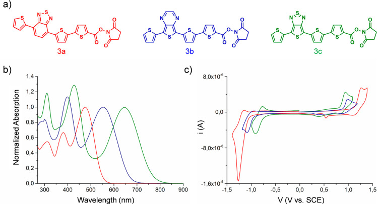
Figure 1.
(a) Molecular structures of the oligothiophene N-hydroxysuccinimidyl esters 3a, 3b, 3c. (b) UV–vis spectra of the compounds 3a (red line), 3b (blue line), 3c (green line) in DMF. The spectra are normalized to the absorption band relative to the lowest energy transition of each molecule. (c) Voltammograms of 3a (red line), 3b (blue line), 3c (green line) in CH2Cl20.1 mol·L–1 (C4H9)4NClO4 on Pt disk electrode (1 mm diameter, scan rate 0.1 v s–1).

