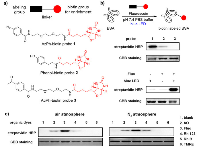
Figure 2.
Photocatalytic labeling of bovine serum albumin (BSA) in vitro. (a) Structures of probes 1–3. The chemical labeling groups and linkers are shown in black, and the biotin groups for enrichment are shown in red. (b) Photocatalytic BSA labeling with biotin probes 1–3 using fluorescein (Fluo) under the blue LED irradiation (468 nm, 5.8 mW/cm2) at 25 °C for 60 min. Control experiments with AzPh-biotin probe 1 showed the fluorescein and the light irradiation were critical for the reaction. The biotinylated-BSA was analyzed by HRP-conjugated streptavidin (streptavidin-HRP), and the BSA proteins were detected by Coomassie brilliant blue staining (CBB staining). BSA (2 μM in pH 7.4 PBS buffer), fluorescein (100 μM), and probes 1–3 (100 μM). (c) Photocatalytic BSA labeling with AzPh-biotin probe 1 (100 μM) by different organic dyes (100 μM) in the air atmosphere or the nitrogen atmosphere under a Xe lamp source at 25 °C for 1 min (15 cm from Xe lamp, 19.8 mW/cm2 with 500 nm band-pass filter for lanes 1–4, 550 nm band pass filter for lanes 5 and 6).

