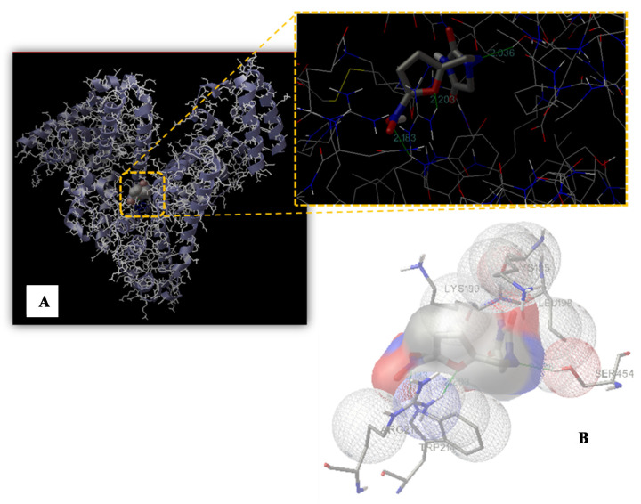
Figure 6.
Binding site of nitrofurantoin to HSA. (A) The HSA structure is rendered with ribbons and lines, while nitrofurantoin is rendered as space fill. The inset shows a magnification of the binding site with nitrofurantoin represented using a stick model. The hydrogen bonds between the ligand and the protein are shown in green and the distance is expressed in Å. (B) Representation of the amino acid residues in the binding site with their van der Waals radii.

