Figure 2.
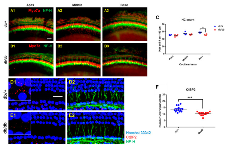
Hair cell loss and synaptopathy in the cochlea of diabetic mice. (A–C) Whole mounts of the auditory epithelium from db/+ and db/db at 14 weeks of age. Tissues were visualized by myosin VIIa (red, hair cells) and NF-H (green, neurofilaments), and photographed using a fluorescence microscope (A,B). Hair cells from apex and middle turns of the cochlea did not show differences between db/+ and db/db mice (C). A significant hair cell loss was found in the base turn of db/db mice compared to db/+ animals (C). n = 4. (D–F) Loss of nerve fibers and synaptic ribbons in diabetic mice at 14 weeks of age. (D,E) Whole-mounts of auditory epithelium were triple-stained with CtBP2 (red, a marker of synaptic ribbons), NF-H (green, neuronal cell marker), and Hoechst (blue, nuclear marker) to evaluate synaptopathy. Severe synaptic loss ((D1) vs. (E1), inset, red) and decreased nerve fibers (green) were observed in the db/db mice compared to the db/+. Number of pre-synaptic marker (CtBP2, red) per 10 hair cells (blue) was quantified (F). Diabetic mice (db/db mice) showed a significant decrease in CtBP2 counts. n = 11. * p < 0.05, *** p < 0.001. Scale bar = 30 μm. All graphs represent mean ± S.E.M. Unpaired t-test.

