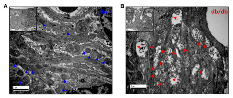
Figure 6.
TEM images of the cochlear stria vascularis (SV). (B) Stria vascularis of db/db mice exerted small vacuolization and gaps between the strial cells. Mitochondria appeared swollen and distorted with reduced cristae in the SV of db/db mice (B), red triangles compared to db/+ animals (A), blue triangles. Scale bar = 2 μm, insert, 5 μm.

