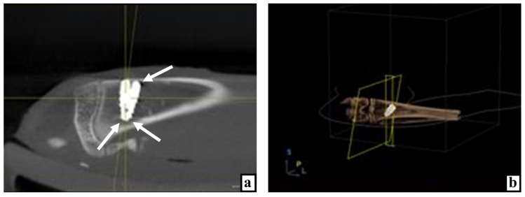
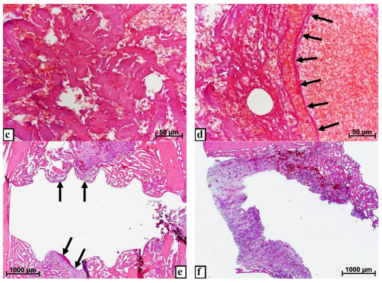
Figure 1.
Results of studying the bone tissue of the proximal tibial condyle in control rabbits at various times after the introduction of dental implants. (a) X-ray image of the implant in the tibia 3 days after surgery, arrows indicate areas of reduced bone density. (b) Computer 3D modeling of the implant position in the bone on the 3rd day after surgery. (c) On the 3rd day after implantation, there are extensive hemorrhages and a large volume of split bone fragments in the cancellous bone, staining with hematoxylin and eosin. (d) Hemorrhages, vascular distention, and congestion (arrows) near and among the split bone particles 3 days after implant placement, stained with hematoxylin and eosin. (e) On the 10th day, the implant mostly borders with the bone tissue and only in small areas it is adjoined by loose fibrous connective tissue (arrows) with a significant number of hyperemic wide thin-walled vessels, stained with hematoxylin and eosin. (f) After 10 days the implant is delimited from the bone throughout its entire length by connective tissue with wide hyperemic vessels, stained with hematoxylin and eosin.


