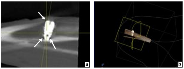
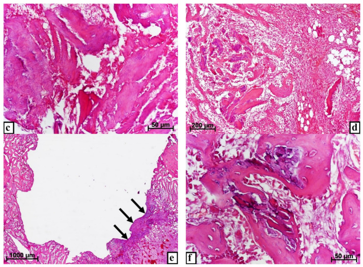
Figure 2.
The state of the animal tibia at various times after the introduction of dental implants into the proximal condyle with the preliminary injection of MSC EVs. (a) By the 3rd day after the operation, according to the X-ray examination, the tissue rarefaction near the implant neck is insignificant (arrow), in the apex area—only along its sides (arrows). (b) Computer 3D modeling of the device position in the bone 3 days after implantation. (c) Three days after the operation, the fragments of bone detritus adhere tightly to each other, between them there are practically no corpuscles and blood vessels, stained with hematoxylin and eosin. (d) On the 7th day after implantation, next to the split parts of the bone tissue infiltrated by large cells, there are structures of new bone, possibly formed from these bone fragments, stained with hematoxylin and eosin. (e) By the 10th day, only for a short distance, the implant borders with the connective tissue (arrows), stained with hematoxylin and eosin. (f) Partial lysis, consolidation with each other, and ingrowth of split bone fragments into new forming bone structures 10 days after surgery, stained with hematoxylin and eosin.


