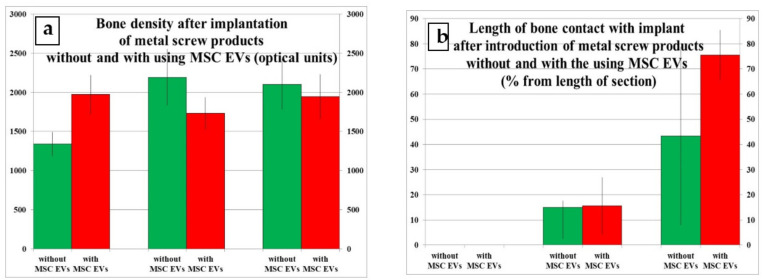
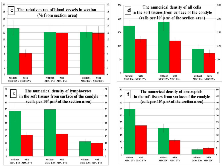
Figure 3.
Results of comparing bone density, vascularization, and leukocyte infiltration in tissues of the rabbit’s proximal tibial condyle at different times after the introduction of screw implants without and with the use of cells. (a) Bone density (optical units) after implantation of metal screw products without and with using MSC EVs. (b) Length of bone contact with implant (% from length of section) after introduction of metal screw products without and with the using MSC EVs. (c) Relative area of blood vessels (% from section area) in section. (d) Numerical density of all cells (per 105 μm2 of the section area) in the soft tissues from surface of the condyle. (e) Numerical density of lymphocytes (per 105 μm2 of the section area) in the soft tissues from surface of the condyle. (f) Numerical density of neutrophils (per 105 μm2 of the section area) in the soft tissues from surface of the condyle.


