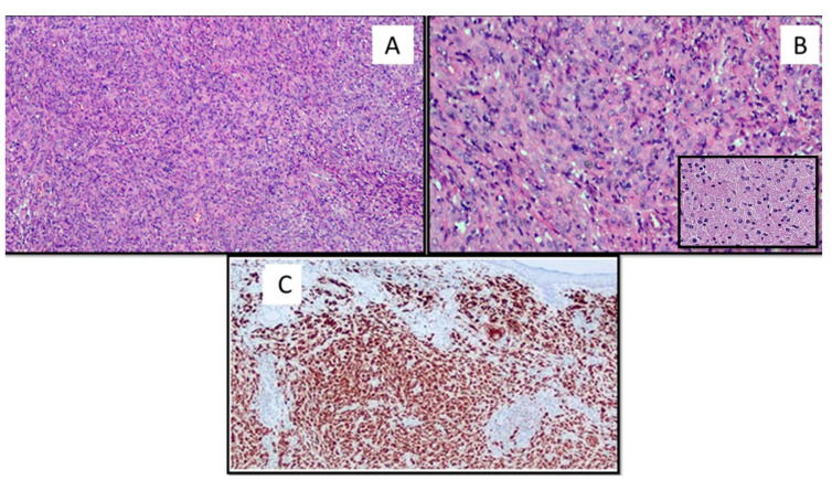
Figure 2.
(A) The lesion was composed of a variable admixture of fibroblast-like cells and histiocytes that showed in more than 85% a large eosinophilic cytoplasm filled of granules or microvacuoles (hematoxylin and eosin, original magnification 40×). (B) Cytological details of the lesion with granular cytoplasm (original magnification 60×). Box: Histological details of the granular cytoplasm of the GCD constituent cells (original magnification 60×). (C) The neoplastic cells were strongly immunoreactive for CD68 (original magnification 40×).

