Figure 1.
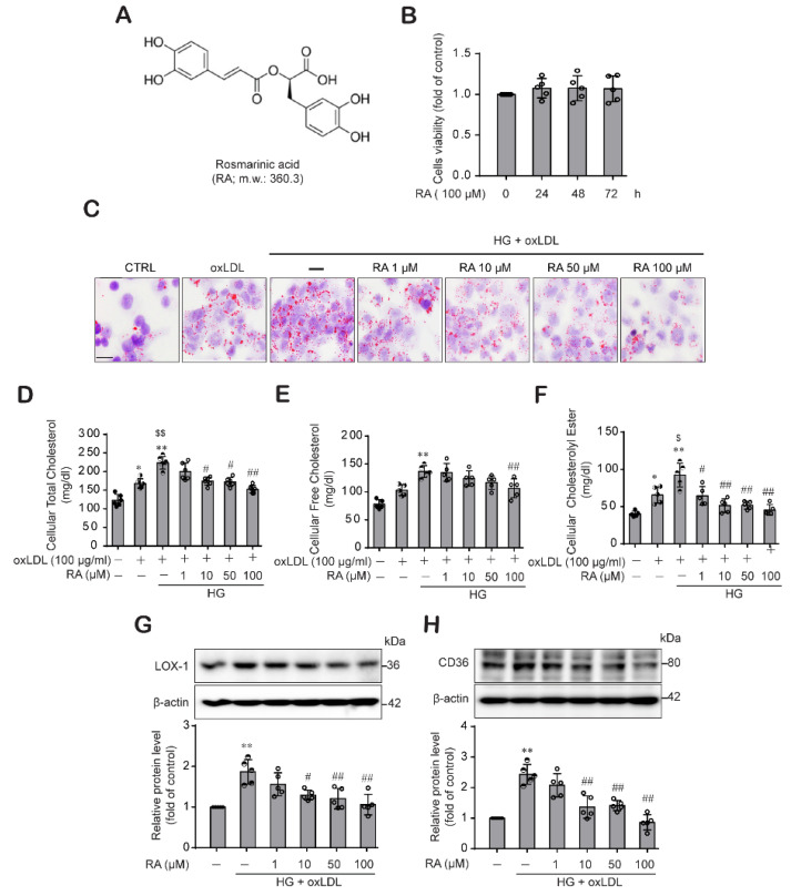
RA significantly reduced the oxLDL-induced lipid contents, cholesterol contents and levels of the SRs LOX-1 and CD36 in macrophage under HG conditions. (A) Chemical structure of RA. (B) Effect of RA on THP-1 macrophage viability. THP-1 macrophages were treated with RA (100 μM) for 24, 48 and 72 h, and then cell viability was measured by the CCK-8 assay as described in the Methods section. Results are presented as the mean ± SD from five independent experiments. (C) THP-1 macrophages were treated with oxLDL (100 μg/mL) under low glucose (5 mM) or HG (25 mM) conditions in the presence or absence of RA (1, 10, 50 and 100 µM). After 24 h, intracellular lipids were stained with Oil Red O (ORO) and observed under light microscopy (400× magnification) (scale bar: 50 μm). Results were confirmed by repeated experiments. (D–F) Cells were treated as described above, and intracellular total cholesterol (D), free cholesterol (E) and cholesteryl ester (F) were measured with a cholesterol/cholesteryl ester quantitation kit according to the manufacturer’s instructions as described in the Methods section. LOX-1 (G) and CD36 (H) protein levels were analyzed from the cell lysates by western blot analysis. The data are presented as the mean ± SD of five independent experiments. * p < 0.05, ** p < 0.01 compared to the control; $ p < 0.05, $$ p < 0.01 compared to the oxLDL; # p < 0.05, ## p < 0.01 compared to the HG + oxLDL group.

