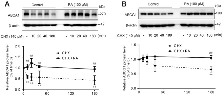
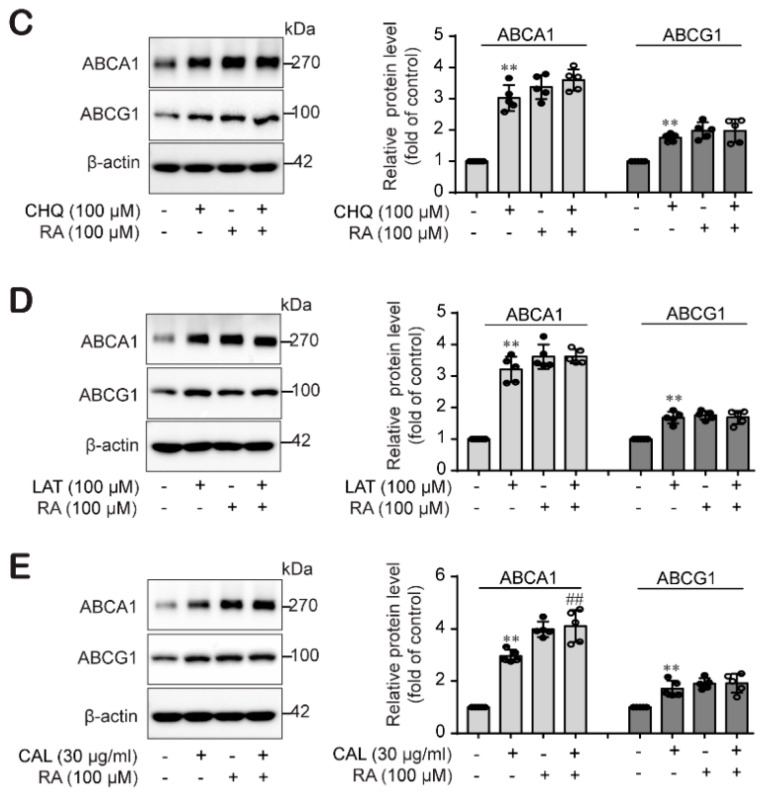
Figure 6.
RA stabilizes ABCA1 protein levels by impairing protein degradation. THP-1 macrophages were incubated for 24 h with or without RA (100 µM) and treated with CHX (140 µM) for different durations (0, 10, 20, 40 and 180 min). Cell lysates were collected to analyze ABCA1 (A) and ABCG1 protein levels (B) by western blot analysis. Band densities were quantified, and relative protein levels are presented as the mean ± SD of five independent experiments. Significance is indicated by ** p < 0.01 compared with the control; ## p < 0.01 compared to the CHK-treated group. (C–E) Cells were treated with or without RA at 100 µM for 24 h and incubated for another 3 h with the lysosomal inhibitor chloroquine (CHQ, 100 µM) (C), the proteasome inhibitor lactacystin (LAT, 10 µM) (D), or the calpain inhibitor calpeptin (CAL, 30 µg/mL) (E). Cell lysates were collected for ABCA1 and ABCG1 protein analysis using western blot analysis. Band densities were quantified, and relative protein levels are presented as the mean ± SD of five independent experiments. ** p < 0.01 compared to the control; ## p < 0.01 compared to the CAL-treated group.


