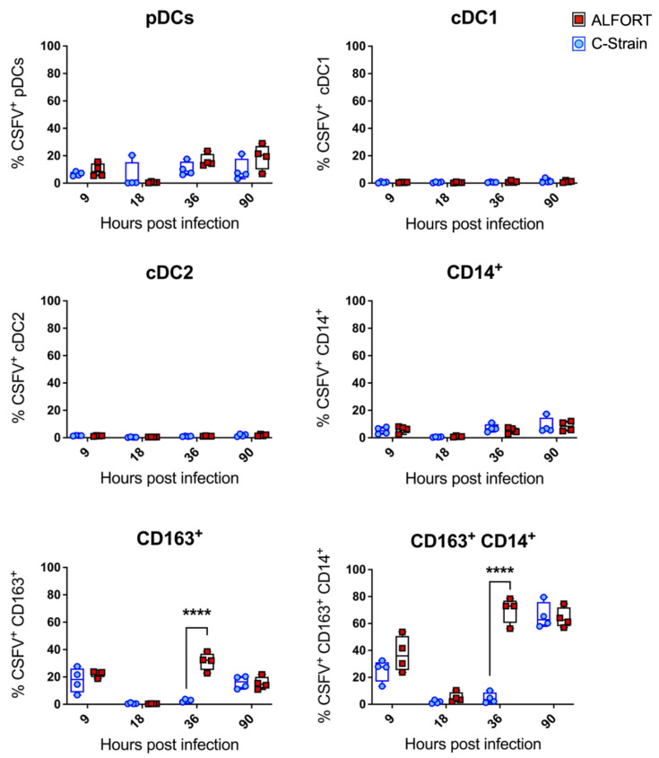
Figure 4.
CSFV infection of tonsillar myeloid cell populations. Tonsillar myeloid cells were stained with antibodies to CD4, CD14, CD172a, CD163, CADM1 and MHC-II followed by intracellular staining for CSFV E2 using the monoclonal antibody WH303 to determine the infection status of each population at each time point. For each time point the median percentage of infected cells for the 4 pigs culled and data are shown. Boxes represent the 25/75% percentile. Statistical significances between the C–strain (blue circles) and Alfort-187 (red squares) were evaluated by a two-way ANOVA with Bonferroni correction (**** p < 0.0001).

