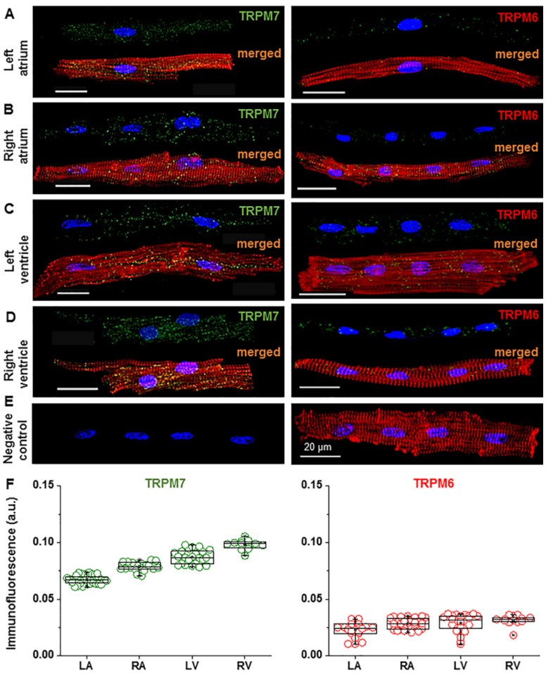
Figure 1.
Immunofluorescence images suggesting the presence of TRPM6 and TRPM7 proteins in pig cardiomyocytes from different cardiac chamber walls. (A–D) Immunofluorescence of TRPM7 (left) and TRPM6 (right) in the left atrium (LA), right atrium (RA), left ventricle (LV), and right ventricle (RV) cardiomyocytes when using Alexa Fluor 488 for the TRPM7 and TRPM6 proteins (stained in green), Alexa Fluor 546 for the F-actin cytoskeleton (stained in red), and Hoechst 33342 for the nuclei (stained in blue). Scale bars indicate 20 µm. (E) Example of a negative control, where the primary antibody for TRPM6 and/or TRPM7 is not added, but the cardiomyocyte was subjected to Hoechst 33342 and Alexa Fluor 546. Under such conditions, only immunofluorescence of the nuclei (stained in blue) and F actin cytoskeleton (stained in red) is detected. Note: same cardiomyocyte in the left and right (merged image) panels (F) Quantification of the staining intensity of the immunodetected fluorescence of TRPM7 and TRPM6 in the four cardiac chamber walls: LA, RA, LV, and RV. The mean data is provided in arbitrary units (a.u.) (see Table 1). A blinded study design (with the origin or treatment of cells unknown to the investigator) was used for the detection of immunofluorescence during the various experimental conditions.

