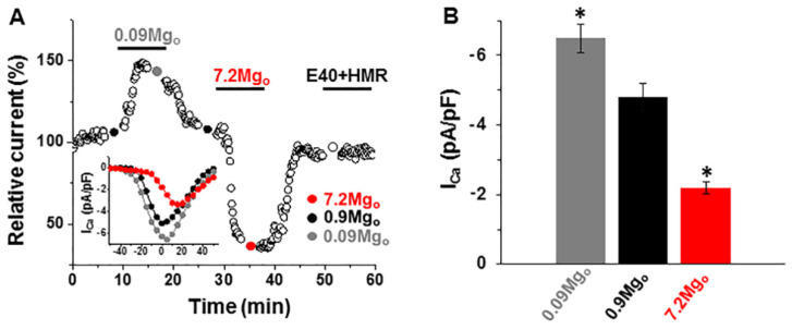
Figure 4.
Effect of changing the external [Mg2+] on the L-type Ca2+ currents (ICa-L). (A) Time diary of the amplitude of the L-type Ca2+ currents obtained by using depolarizing steps for various potentials in a cell dialyzed with 0.8-mM [Mg2+] and superfused with extracellular solutions containing 0.09-mM, 0.9-mM, or 7.2-mM [Mg2+]. Notice the increase vs. suppression of the ICa-L amplitude by low vs. high [Mg2+]o and the lack of effect of the IK inhibitors (E4031/HMR1556). Bottom inset: The current–voltage relations obtained by the depolarizing steps to the potentials ranging from −50 mV to +50 mV in the same cell. (B) Summary data of the peak ICa-L amplitude and the effects of lowering and raising the [Mg2+]o. * p < 0.05 vs. 0.9-mM [Mg2+]o (n = 8–13).

