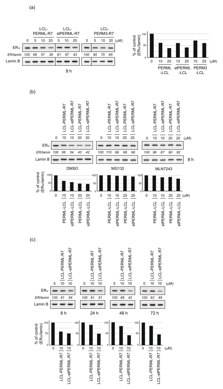
Figure 4.
Degradation of ERα by the synthesized peptides analyzed by quantitative Western Blotting. (a) MCF-7 cells that had been cultured in RPMI 1640 medium containing 10% fetal bovine serum were treated with the indicated concentrations of LCL-PERML-R7, LCL-stPERML-R7 or LCL-PERM3-R7 for 8 h. (b) MCF-7 cells were treated with the indicated concentrations of LCL-PERML-R7 or LCL-stPERML-R7 in the presence or absence of 10 µM MG132 or MLN7243 for 8 h. (c) MCF-7 cells were treated with 10 µM LCL-PERML-R7 or LCL-stPERML-R7 for different times (8, 24, 48 and 72 h). Whole-cell lysates were analyzed by Western blotting with the indicated antibodies. Numbers below the ERα panels represent ER/lamin ratios, normalized by designating the expression from the vehicle control condition as 100%. The changes in protein levels were reproducible between the two independent experiments. Data in the bar graph are the mean of two results.

