Figure 1.
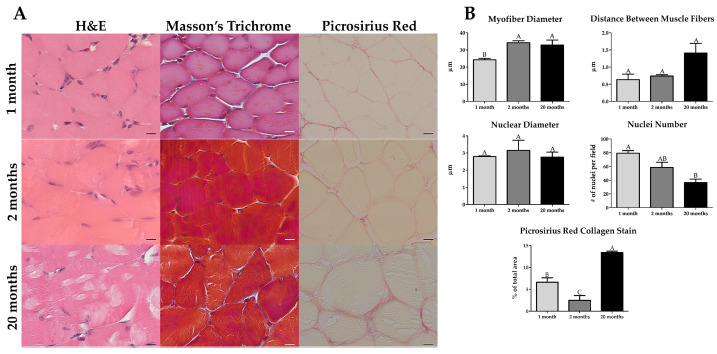
The gastrocnemius shows histomorphometric differences in old age. (A) Hematoxylin and Eosin (H&E) staining (n = 3) revealed a reduction in nuclei and the presence of lipofuscin-like or tubular aggregates inside the muscle fibers with old age. Masson’s Trichrome (n = 3) had no apparent differences with age. Picrosirius Red staining (n = 4) displayed a pronounced increase in collagen staining in old age. (B) When quantified histomorphometrically, the gastrocnemius had a smaller myofiber diameter in immature muscle, an increase in the distance between muscle fibers and a decrease in the number of nuclei in old age, and a bimodal amount of collagen staining where immature and old have higher levels of collagen than young adult, albeit older also has higher amounts than immature muscle. Scale bar = 10 µm. Groups that do not share a letter (e.g., A, B, or C) are statistically different according to a one-way ANOVA followed by a two-tailed Tukey’s correction (p < 0.05, n = 3 for H&E and Masson’s, n = 4 for Picrosirius Red).

