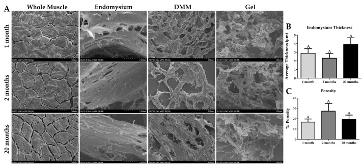
Figure 4.
Scanning Electron Imaging shows distinctions in ECM structure with age in native muscle, decellularized muscle matrix (DMM), and processed DMM gels. (A) The muscle fibers are surrounded by endomysium in the whole muscle images, and the space between the muscle fibers appear to increase with age, however not significantly. DMM (n = 4 for 20-month DMM) and processed DMM gels, there is an obvious sclerotic structure indicating the presence of polymerized ECM such as collagen. (B) Endomysium thickness trends upwards with age but is not significantly different. (C) The porosity of DMM is not affected by age. Groups that do not share a letter (e.g., A) are statistically different according to a one-way ANOVA followed by a two-tailed Tukey’s correction (p < 0.05, n = 6 except the 20-month-old muscle has n = 4).

