Figure 8.
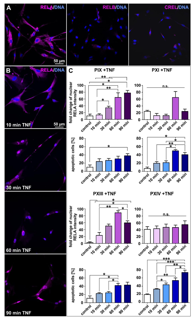
Tumor necrosis factor treatment led to profound cytotoxicity in prostate cancer stem cells unable to activate NF-κB. (A) The exemplary immunostaining of cultured PXIV prostate cancer stem cells (PCSCs) resulted in predominant NF-κB RELA expressions as well as slightly detectable RELB and CREL proteins expressions. (B) Exemplary images of immunocytochemically NF-κB RELA-stained PXIII PCSCs after treatment with tumor necrosis factor alpha (TNFα) for 10, 30, 60 and 90 min. The expressions of RELA in PCSCs seemed to shift from the cytoplasm to nucleus over the experimental duration. Nuclear counterstaining was performed using DAPI. (C) TNFα stimulations were evaluated by measuring the fold changes in nuclear RELA intensities and average percentages of apoptotic cells via quantification with ImageJ software (NIH, Bethesda, MD, USA). The PIX and PXIII PCSC populations displayed statistically significant upregulated fold changes of nuclear RELA intensities upon TNFα treatment (left panel, magenta graphs). The DAPI staining of nuclei displayed statistically high upregulations of apoptotic cells in PXI and PXIV PCSCs after treatment with TNFα (right panel, blue graphs). Statistical analysis was performed using Mann–Whitney U tests (minimum 50 cells per condition and population); * p ≤ 0.05, ** p ≤ 0.01 and *** p ≤ 0.001 were considered significant. Mean ± standard error of the mean (SEM).

