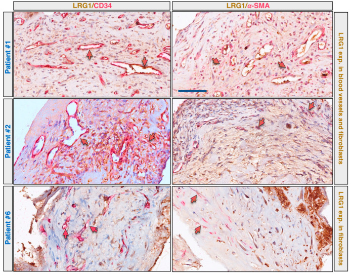
Figure 3.
LRG1 is expressed in pathological vessels and myofibroblasts of CNVM. CNV membrane sections were analyzed by multiplex immunohistochemistry using anti-LRG1 (brown staining) and anti-CD34 (red staining), or anti-LRG1 (brown staining) and anti-α-SMA (red staining) antibody combinations. Results of three representative patients are shown. Red arrows indicate blood vessels negative for LRG1; red/brown arrows indicate blood vessels or myofibroblasts positive for both LRG1 and CD34, or LRG1 and α-SMA. Scale bar: 50 μm.

