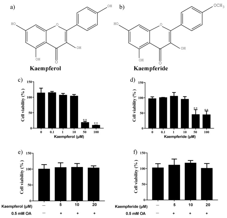
Figure 3.
Changes in viability of HepG2 cells after incubation with kaempferol and kaempferide. (a) Chemical structure of kaempferol. (b) Chemical structure of kaempferide. (c) HepG2 cell viability after incubation with kaempferol. (d) HepG2 cell viability after incubation with kaempferide. (e) No change in HepG2 cell viability by co-incubation of OA and kaempferol for 48 h. (f) No change in HepG2 cell viability by co-incubation of OA and kaempferide for 48 h. Data were expressed as Mean ± SD of three independent experiments (n = 3). ** p < 0.01, compared with vehicle-treated control.

