Figure 5:
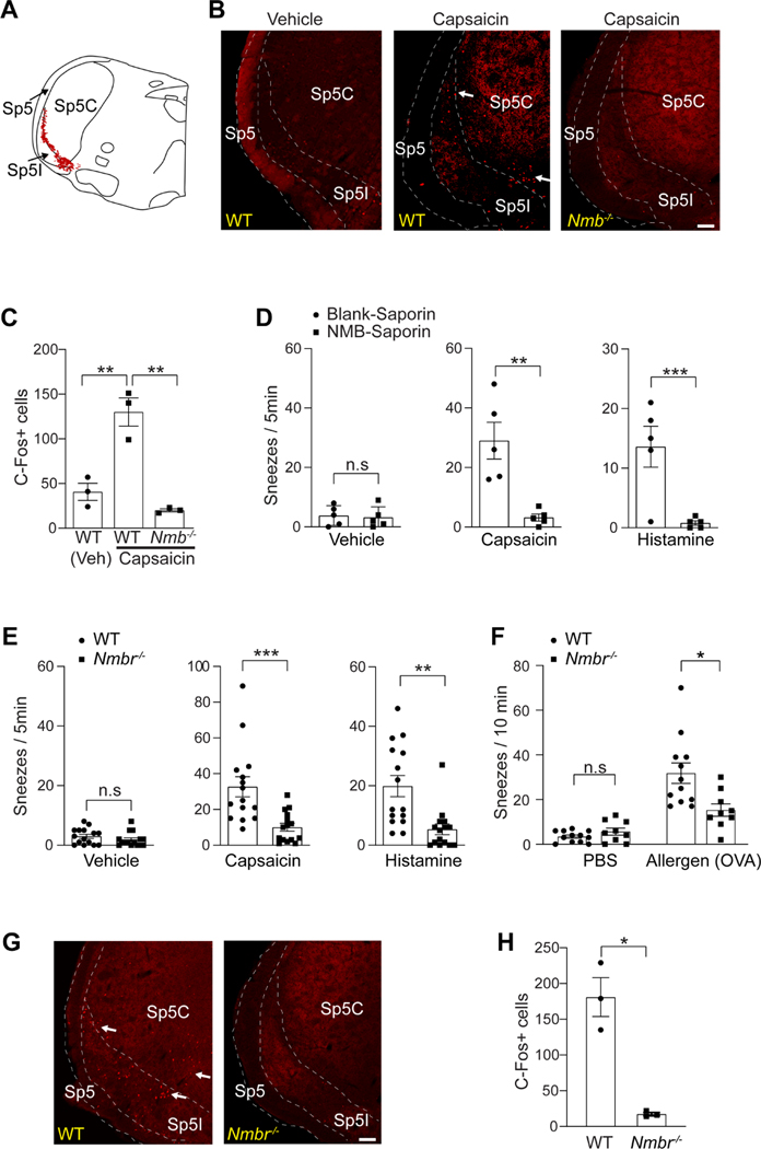
NMB-sensitive postsynaptic neurons in the sneeze-evoking region mediate sneezing. (A) Diagram showing the sneeze-evoking region (i.e., the central projection of nasal sensory neurons; indicated by red dots) in the spinal trigeminal nucleus (SpV) based on previous studies. Sp5, spinal trigeminal tract; Sp5C, spinal trigeminal nucleus caudalis; Sp5I, spinal trigeminal nucleus interpolar. (B) Capsaicin-induced sneezes lead to a significant increase in the expression of c-Fos within the sneeze-evoking region of WT (indicated by arrows), compared with the saline vehicle control. Nmb-deficient mice display a significantly reduced c-Fos signal. (C) Quantification of c-Fos+ neurons in the sneeze-evoking region after saline (Veh) or capsaicin treatments. (D) Microinjection of NMB-saporin into the sneeze-evoking region abolishes the sneezing responses to aerosolized capsaicin (12 μM) and histamine (100 mM) solution. (E-F) Nmbr−/− mice display significantly reduced sneezing responses to aerosolized capsaicin (12 μM), histamine (100 mM) solution and allergen (ovalbumin, OVA, 0.2 μg in 2 μl PBS / nostril) stimuli. (G-H) Capsaicin-induced sneezes lead to the expression of c-Fos in the sneeze-evoking region of WT (indicated by arrows) but not Nmbr−/− mice. All images shown are representatives of three biologically independent mice. Scale bars: 100 μm. The total number of c-Fos+ neurons in the sneeze-evoking region was counted from ten sections per mouse (n=3 / genotype). Each dot represents an individual mouse. n=5–15 mice/group for behavioral tests. Data are represented as mean ± SEM. * P≤0.05, ** P≤0.01, *** P≤0.001, n.s. not significant.

