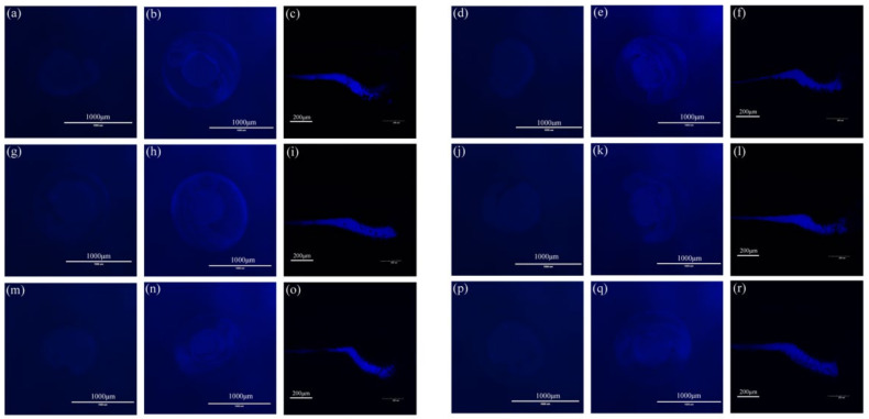
Figure 10.
Fluorescence images of zebrafish embryos after soaking with compounds (a) 1, (d) 2, (g) 3, (j) 4, (m) 5, and (p) 6 (15 µM) for 1 h at 24 hpf. Fluorescence images of zebrafish embryos after soaking with compounds (b) 1, (e) 2, (h) 3, (k) 4, (n) 5, (q) 6 (15 µM) for 1 h at 48 hpf. Fluorescence images of zebrafish embryos after soaking with compounds (c) 1, (f) 2, (i) 3, (l) 4, (o) 5, (r) 6 (15 µM) for 1 h at 120 hpf.

