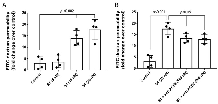
Figure 2.
S1 spike protein alone caused disruption of the pulmonary endothelial cell monolayer. (A) HPAEC were grown on Transwell inserts to form uniform monolayer and then were exposed to various concentrations of S1 spike protein (5–25 nM) for 12 h in the presence of 40 kD-FITC conjugated dextran. Endothelial monolayer integrity was assessed by measuring the passage of FITC-dextran molecule through the monolayer. FITC-green fluorescence was monitored by fluorescence plate reader as described in Section 4. (B) In a similar set of experiments, HPAEC were co-incubated with an anti-ACE2 antibody (100 nM) and S1 spike protein (25 nM) for 12 h in the presence of 40 kD-FITC conjugated dextran. Values are presented as average fold difference over control. (n = 4). Bars represent average mean value, each dot in the bars represent individual data points and vertical error bars represent SD. Unpaired Student’s t-test. Statistical significance between groups were indicated by brackets with p values.

