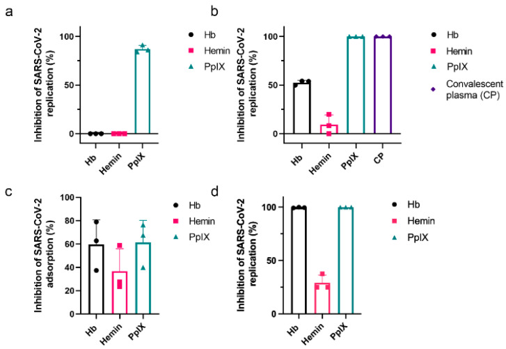
Figure 5.
Inhibition of SARS-CoV-2 replication and attachment by porphyrins and Hb. Vero E6 cells (a) or SARS-CoV-2 (b) were pre-incubated with 1 µM of Hb, hemin, or PpIX for 1 h at 37 °C before the start of infection with an MOI of 0.01 for an additional hour at 37 °C. Culture supernatants were collected after 24 h, and the virus titer was determined by plaque assay (n = 3). A convalescent plasma of an infected patient (1:3 dilution) was used as the positive control. (c) SARS-CoV-2 was incubated at 1 µM Hb, hemin, or PpIX for 1 h at 37 °C before introducing Vero E6 cells at an MOI of 0.01 for 1 h at 4 °C. Rinsed cell monolayers were lysed, and RT-PCR quantified the virus content. Cumulative data (Hb, Hemin, PpIX: n = 3). (d) Vero E6 was infected with SARS-CoV-2 at an MOI of 0.01 for 1 h at 37 °C and then treated with 1 mM of hemin, PpIX, or Hb. After 24 h, supernatants were collected, and the virus titer was quantified by PFU/mL (Hb, Hemin, PpIX: n = 3). Data represented the percentage of inhibition compared to control (infected and untreated) and expressed as mean with standard deviation. Source data are provided as a Source Data file.

