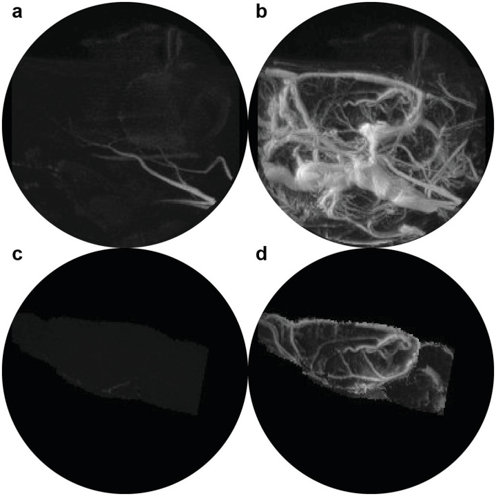
Fig 1. QUTE-CE MRI angiograms.
Maximum intensity projection images (MIPs) of the whole rat head at 8 months are displayed (a) pre-contrast and (b) post-contrast 14mg/kg ferumoxytol. Unique contrast-enhanced vascular MIPs are obtained, and segmentation of the brain from the pre- and post-contrast images (c and d, respectively) demonstrates that time-of-flight signal enhancement is limited to the arteries at the periphery of the field of view and so the signal does not contribute to biomarker measurements.

