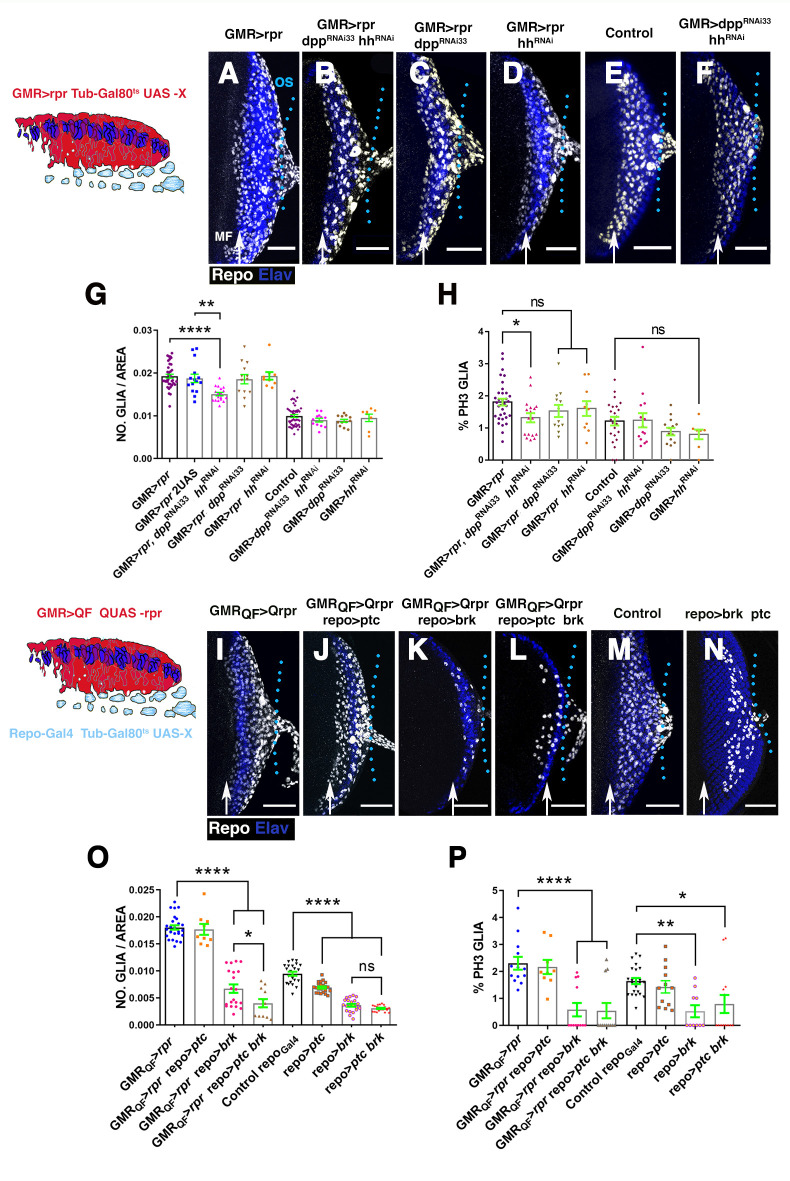
Fig 8. The down-regulation of Dpp and Hh signalling reduce glial response.
(A–F and I–N) Projections of confocal images of third instar eye discs stained with anti-Elav (blue) and anti-Repo (white). (A–F) Effects caused by the down-regulation of Dpp and Hh signalling in the damaged region. The schematic illustration on the left represents a transverse section of an eye disc where the region marked in red (expression domain of GMR-Gal4) corresponds to the area of the discs that has been damaged at the same time that Dpp and/or Hh signalling were depleted. (A) UAS-rpr/+; GMR-Gal4 tub-Gal80ts/+ damaged eye disc. (B) Damaged UAS-rpr/+; GMR-Gal4 tub-Gal80ts/ UAS-hhRNAi; UAS-dppRNA33i. (C) Damaged UAS-rpr/+; GMR-Gal4 tub-Gal80ts; UAS-dppRNAi33 (BDSC33618). (D) UAS-rpr/+; GMR-Gal4 tub-Gal80ts/ UAS-hhRNAi damaged discs. (E) Control undamaged GMR-Gal4 tub-Gal80ts/+ disc. (F) GMR-Gal4 tub-Gal80ts/ UAS-hhRNAi; UAS-dppRNAi33 eye discs. (G and H) Graphs show glial cell density (G) and percentage of mitotic glia cells of discs shown in A–F. (I–N) Effects of the down-regulation of Dpp and Hh signalling in glial cells in damaged discs. In the schematic illustration on the left is indicated in red the region that has been damaged (GMR-QF; QUAS-rpr) and in light blue glial cells. (I) GMR-QF; tub-Gal80ts; repo-Gal4 QUAS-rpr eye disc. (J) GMR-QF; tub-Gal80ts/UAS-ptc; repo-Gal4 QUAS-rpr. (K) GMR-QF; tub-Gal80ts; repo-Gal4 QUAS-rpr/UAS-brk. (L) GMR-QF; tub-Gal80ts/UAS-ptc; repo-Gal4 QUAS-rpr/UAS-brk. (M) Control undamaged tub-Gal80ts; repo-Gal4 disc. (N) tub-Gal80ts/UAS-ptc; repo-Gal4 /UAS-brk. (O and P) Graphs show glial cell density (O) and percentage of mitotic glial cells (P) of discs shown in I–N. Scale bars, 50 μm. Statistical analysis is shown in Table G in S1 Text. The numerical data used in this figure are included in S1 Data. GMR, glass multiple reporter; Dpp, Decapentaplegic; Hh, Hedgehog; RNAi, RNA interference; rpr, reaper.

