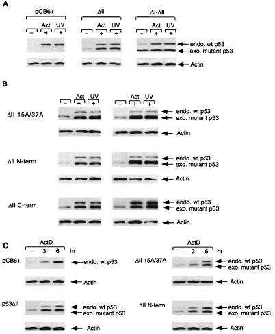
FIG. 7.
Stable expression of p53ΔII combination mutant proteins in MCF-7 cells which express wild-type (wt) p53. Stable clones were isolated, and p53 protein levels were assessed by Western blot analysis using PAb1801 or D01 after treatment with either 5 nM actinomycin D (Act; harvested at 10 h) or 50-J/m2 UV (harvested at 6 h). (A) Western blot analysis of MCF-7 stable cell lines expressing either the pCB6+ control (leftmost blot), p53ΔII (middle blot), or p53ΔI-ΔII (rightmost blot). Equal protein loading was confirmed by actin expression. endo., endogenous; exo, exogenous. (B) Western blot analysis of two independent MCF-7 stable cell lines (clone 1, left; clone 2, right) expressing either p53ΔII15A/37A (top), p53ΔIIN-term (middle), or p53ΔIIC-term (bottom). Equal protein loading was confirmed by actin expression. (C) Western blot time course analysis of actinomycin D (ActD) treatment (0, 3, and 6 h) of MCF-7 cells stably expressing either pCB6+ (control), p53ΔII (clone 1), p53ΔII15A/37A (clone 1), and p53ΔIIN-term (clone 1). All blots were reprobed with an antiactin monoclonal antibody to assess protein levels. Equal protein loading was confirmed by actin expression.

