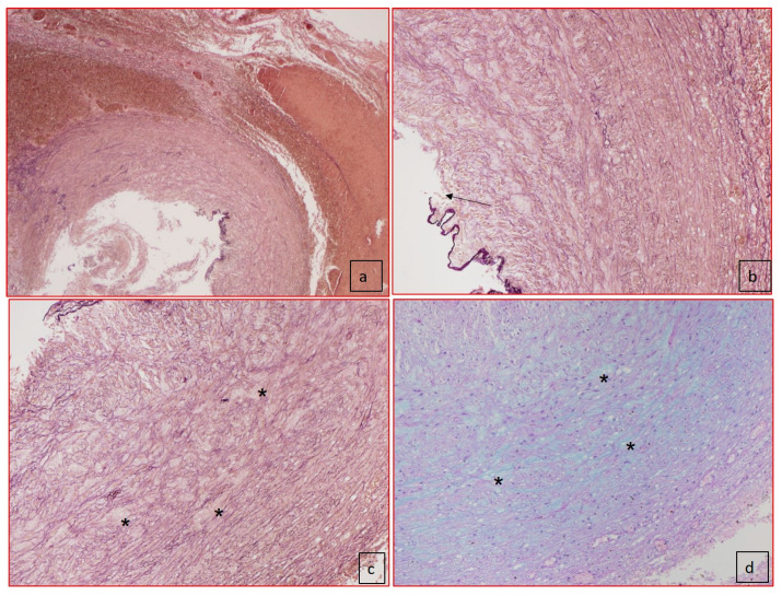
Figure 3.
Histology of the ductus arteriosus (case 2). (a) Panoramic view of ductus arteriosus with elastic fiber–van Gieson staining showed the dissection of media (50× of magnification) and the absence of intimal cushions. (b) Elastic fiber–van Gieson staining at higher magnification showed the disorganization of elastic fibers and rupture of internal elastic lamina (black arrow). (c) Elastic fiber–van Gieson (100× magnification) staining showing disorganization of elastic fibers and presence of lacunar spaces, cystic medial necrosis (*), without SMCs replaced by mucopolysaccharides as shown in (d) with Alcian–PAS staining (100× magnification).

