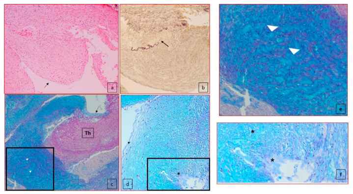
Figure 4.
Histology of the ductus arteriosus. (a) Subendothelial intimal cushion (case 5) (50× magnification, black arrow); (b) elastic fiber–van Gieson (50× magnification) showed the interruption of the internal elastic lamina (black arrow); (c) presence of lacunar spaces (white arrow head) in the media closed to dissection (black arrow) (100× magnification) with thrombus formation (Th); (d) lacunar spaces, cystic medial necrosis (*) in the media, under the intimal proliferation (black arrow head) (100×). (e) High-power view of the insert highlighted in (c). (f) High-power view of the insert highlighted in (d).

