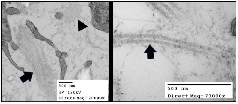
Figure 2.
TEMs of the collagen fibers in in Hamburger Hamilton (HH) stage 42 chick (Gallus gallus) valve leaflets. The image in the right panel shows bundles of collagen fibrils arranged in two distinct originations. The fiber oriented in the plane of the figure (black arrow) displays the “D-period” striations. The fiber oriented into the plane of the figure (black arrowhead) shows the quasi-hexagonal arrangement of collagen fibers in cross-section.

