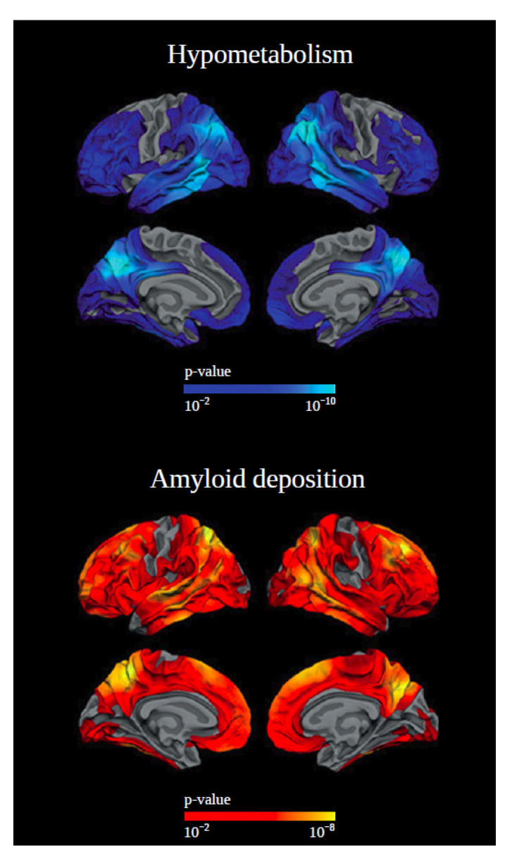
Figure 1.
Example of positron emission tomography (PET) scans from individuals with Down syndrome (DS) with symptomatic Alzheimer’s disease (AD) compared with those with no clinical evidence of dementia. The upper panel represents a decrease in brain glucose metabolism of the temporoparietal, precuneus-posterior cingulate, and frontal brain regions in symptomatic DS. The lower panel shows the increased global cerebral Aβ deposition, with a relative sparing in sensory and motor areas in DS with symptomatic AD. Adapted with permission from [13].

