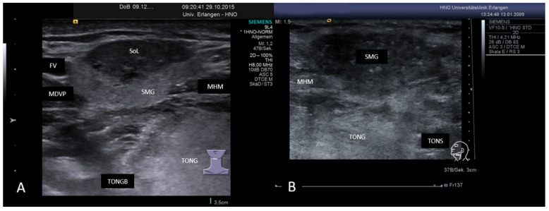
Figure 17.
(A,B) Sclerosing IgG-4-related sialadenitis in two SMGs. In (A), the caudal part of the right-sided SMG is characterized by a mixed hypoechoic to anechoic and hyperechoic tissue pattern. In (B), the whole gland is changed in this way. A functioning parenchyma is no longer visible. Clinical diagnosis was first presence of a “Küttner’s tumor”. Abbreviations: SMG, submandibular gland; MHM, mylohyoid muscle; MDVP, digastric muscle venter posterior; TONG, tongue; TONGB, base of tongue; TONS, tonsil; FV, facial vein; SoL, space occupying lesion.

