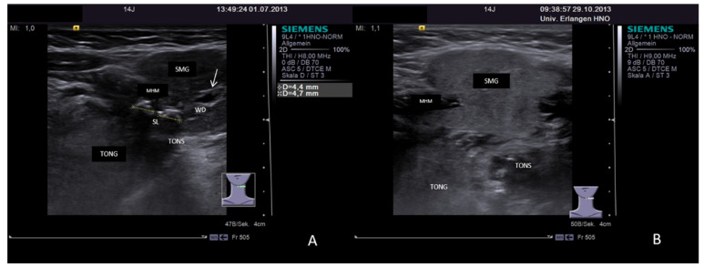
Figure 19.
(A,B) Case of sialolithiasis of left-sided SMG in a 14-year-old patient demonstrating post-treatment recovery of the gland. Pre-treatment status shows two sialoliths within the hilum of the gland measuring 4.4 and 4.7 mm. The gland parenchyma is enlarged due to congestion with central duct dilation (white arrow, A). Post-treatment US nearly 4 months later demonstrates recovery of the gland. The parenchyma turned out to a have a nearly normal, slightly hyperechoic tissue texture, and no duct obstruction, and congestion is no longer visible (B). Abbreviations: SMG, submandibular gland; MHM, mylohyoid muscle; TONG, tongue; TONS, tonsil; SL, sialolithiasis.

