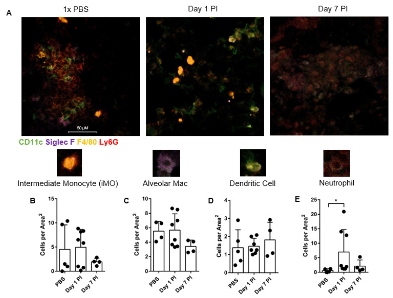
Figure 4.
Immunohistochemistry and lung image scoring reveals heightened CD11c positive and Ly6G positive cells in the lung post infection. Lungs were processed at day 1 and day 7 post infection (PI) and fresh-frozen. (A) In the representative plot, PBS is the mock infection whereas Day 1 and Day 7 show 105 arthroconidia intranasal infection at 40× confocal imaging. 50 μM scale bar applies to all images. (B) Intermediate monocytes (iMO) are defined as F4/80+ Ly6G+, (C) alveolar macrophages (Mac) as F4/80+ CD11c+ Siglec-F+, (D) DCs as CD11c+ F4/80−, and (E) neutrophils as Ly6G+ F4/80− CD11c−. Images scored are from 40× magnification. Each image represents 742,819.8969 uM2 in Area2, calculated from square image of 862 × 862 μm2. Images were blind scored by two independent counters. For all plots displayed line indicates mean, and each dot is one experimental replicate for each lung sample averaged over 5 images and two blind-scorers. n = 4–7, representative of 4 experiments. Comparisons were made between mock PBS infection and infected at each time point. Data was analyzed using unpaired Student’s t-test and outliers excluded using Grubbs Outlier exclusion analysis; * p < 0.05.

