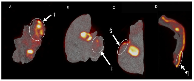
Figure 3.
The 18F-FDG uptake in pathologically proven non-malignant regions. The images display regions of increased 18F-FDG uptake that were later pathologically proven to be non-malignant tissue. (A) displays the coronal view of the resected squamous cell carcinoma (SCC) of the floor of mouth in patient 1. Here, we see an increased uptake in a large region that was later shown to be highly active non-malignant inflamed tissue (†). (B,C) display the coronal and sagittal view, respectively, of the hemiglossectomy for a mucosal SCC in patient 2. Here, we see an increased uptake of a salivary gland (‡) and premalignant dysplastic tissue on the tongue’s epithelia (§). (D) displays the imaging results found after imaging a nasal resection specimen for an angiosarcoma of the nasal tip in patient 6. Here, ¶ displays an increased uptake in the subcutaneous nasal sebaceous glands.

