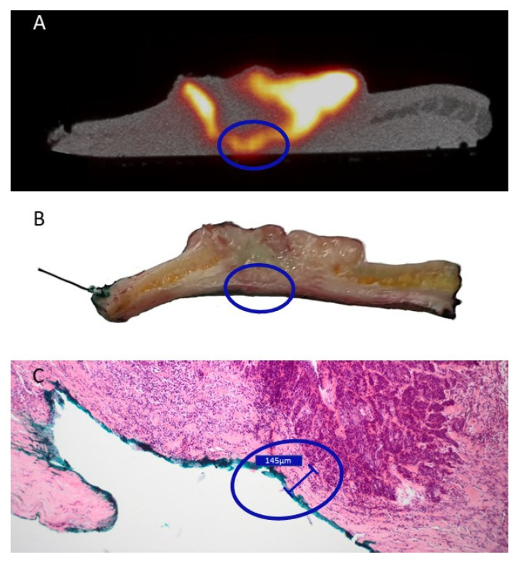
Figure 4.
Correlation between the close margin as described during pathological assessment and PET/CT-imaging of a cutaneous squamous cell carcinoma of the scalp in patient 3. The blue circle encircles a region of interest that showed positive margin on PET/CT-imaging (A), ambiguous margin on the macroscopic pathological assessment (B), but was found to be a ‘close’ margin of 0.15 mm on final microscopical analysis (C). (A): Transversal view of PET/CT imaging of the resected specimen. (B): The sliced tissue of the proposed region corresponding with the PET/CT-imaging found in (A). (C): microscopic pathological assessment of the hematoxylin and eosin-stained slide made after fixation of the macroscopic slide displayed in (B).

