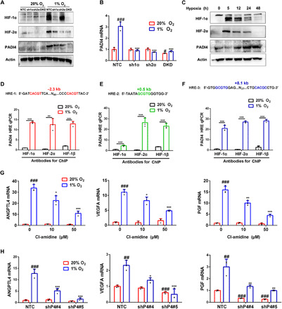
Fig. 1. Hypoxia-induced PADI4 expression is HIF-dependent and is required for HIF target gene transcription.

(A) SUM159 subclones expressing a nontargeting control (NTC) shRNA, an shRNA targeting HIF-1α (sh1α) or HIF-2α (sh2α), or shRNAs targeting both HIF-1α and HIF-2α [double knockdown (DKD)] were exposed to 20 or 1% O2 for 6 hours (HIF-1α and HIF-2α) or 48 hours (PADI4 and actin), and immunoblot (IB) assays of whole-cell lysates (WCLs) were performed. (B) SUM159 subclones were exposed to 20 or 1% O2 for 24 hours followed by RT-qPCR (mean ± SD; n = 3). #P < 0.05, ###P < 0.001 versus NTC at 20% O2; ***P < 0.001 versus NTC at 1% O2 [two-way analysis of variance (ANOVA)]. (C) SUM159 cells were exposed to 1% O2, and IB assays were performed. (D to F) SUM159 cells were exposed to 20 or 1% O2 for 16 hours, and ChIP assays were performed. Primers encompassing HIF binding sites located 2.3 kb 5′ (D), 0.5 kb 3′ (E), and 8.1 kb 3′ (F) to the PADI4 TSS were used for qPCR. Results were normalized to the first lane (mean ± SD; n = 3). **P < 0.01, ***P < 0.001 versus 20% O2 (Student’s t test). (G) SUM159 cells were exposed to 20 or 1% O2 for 24 hours followed by RT-qPCR. Results were normalized to lane 1 (mean ± SD; n = 3). #P < 0.05, ##P < 0.01, ###P < 0.001 versus vehicle at 20% O2. **P < 0.01, ***P < 0.001 versus vehicle at 1% O2 (two-way ANOVA). (H) SUM159 subclones were exposed to 20 or 1% O2 for 24 hours followed by RT-qPCR (mean ± SD; n = 3). ##P < 0.01, ###P < 0.001 versus NTC at 20% O2. *P < 0.05, **P < 0.01, ***P < 0.001 versus NTC at 1% O2 (two-way ANOVA).
