
Figure 1. Solution structure of E.coli NusB.
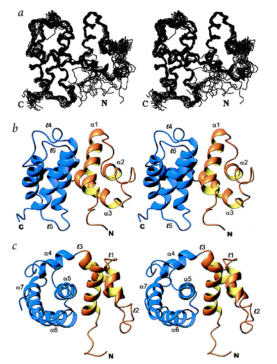
a, Stereo view showing the superposition of 15 of the lowest energy structures of NusB. b, Stereo view of a ribbon trace of NusB. The N-terminal subdomain is colored gold, and the C-terminal subdomain is colored purple. Helices and loops are labeled as discussed in the text. c, Stereo view showing a 90y° rotation of (b).