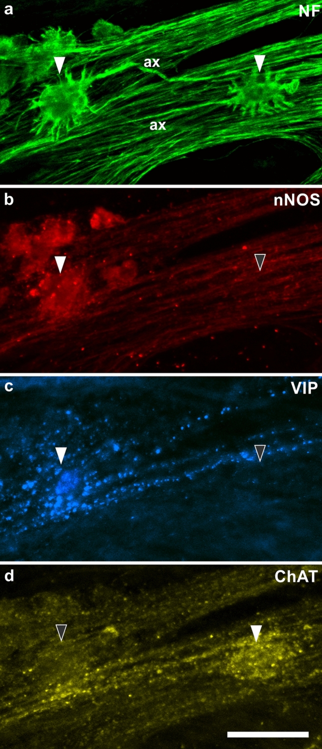
Fig. 2.

a Two “Dogiel type I” neurons (filled arrowheads) stained for neurofilaments (NF). The left one is a spiny neuron, its axon (ax) runs to the right (i.e. anally); the right one is a stubby neuron with an axon (ax) running to the left (i.e. orally). b Corresponding demonstration of staining for neuronal nitric oxide synthase (nNOS). The spiny neuron is positive, the stubby one negative (filled and empty arrowhead, respectively). c The spiny neuron is co-reactive for vasoactive intestinal peptide (VIP), the stubby neuron is negative (filled vs. empty arrowhead). d The spiny neuron is negative for choline acetyltransferase (ChAT), the stubby neuron is positive (empty vs. filled arrowhead). (From ascending colon of a 104-year-old woman; body donated to the Institute of Anatomy) Bar = 50 µm. (Antibodies: NF: Sigma-Merck N0142; nNOS: Novus Biologicals NB120-3511; VIP: Dianova T-5030; ChAT: Merck-Millipore AB144P)
