Fig. 1.
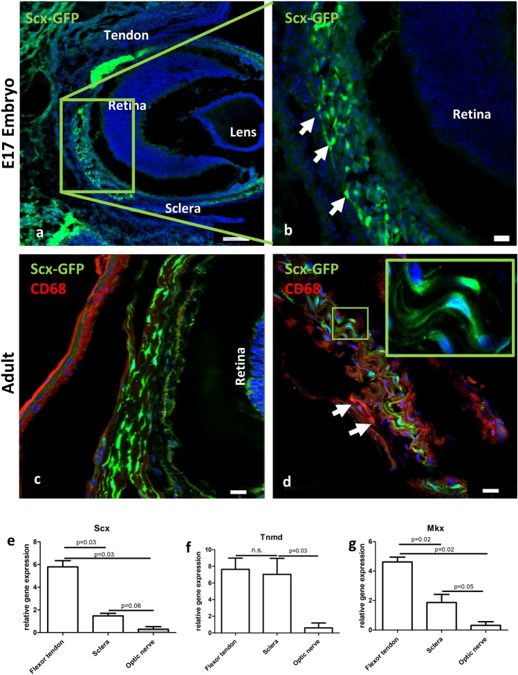
Scleral cells express tendon-associated markers. a, b Fluorescence microscopy revealed numerous scleraxis-GFP (Scx-GFP, green) positive cells in the sclera of embryonic mice at E17. b represents a magnification of the boxed area in a, highlighting the Scx-GFP positive cells (arrows). Blue = DAPI. c, d A similar situation was found in adult mice: spindle shaped Scx-GFP positive cells (green) were present throughout the entire sclera. Immunohistochemistry with CD68 revealed absence of co-localization with the Scx-GFP signal (c), while CD68-immunoreactivity was detected in few cells within the sclera (d, arrows). Blue = DAPI Scale bars: a = 100 µm, b–d = 20 µm. qRT-PCR analysis shows the expression of mRNA of the tendon-associated markers scleraxis (e), tenomodulin (Tnmd, f) and mohawk (Mkx, g), in both tendon and sclera. To a significantly lesser extent, expression is also detectable in the optic nerve.

