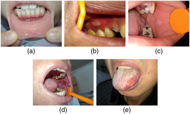
Fig. 3.
Clinical features of sample images in the five categories. (a) Normal mucosa image from lower labial mucosa and upper lip of a healthy subject. (b) Aphthous ulcer on the attached gingiva. (c) A homogeneous white patch on the left buccal mucosa. (d) An extensive white lesion with a red component affecting the whole left buccal mucosa. (e) A malignant lesion on the lateral border of the tongue.

